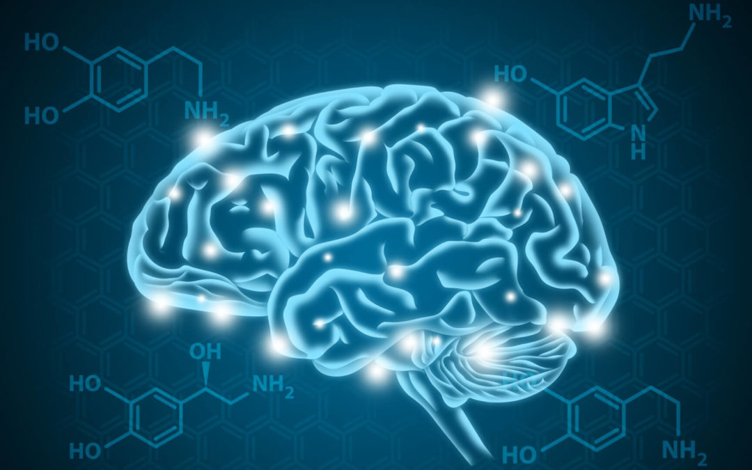
In humans, the TAAR1 (Trace amine-associated receptor 1) gene is responsible for the regulation of amine-associated protein. This protein is mainly expressed in various peripheral organs and cells, such as the small intestine, stomach, white blood cells, and astrocytes. It also interacts with the presynaptic plasma membrane and the intracellular milieu.
The discovery of the TAAR1 gene in 2001 was made by two groups of investigators. These investigators were able to identify the six functional amine-associated receptors that are known to bind to endogenous amines. It is known that this protein controls the neurotransmission of certain chemicals in the central nervous system (CNS). It also affects the function of the neuroimmune system.
The role of TAAR1 in the regulation of energy metabolism has received relatively little attention. It is possible that this function is mediated by either central or peripheral effects.
The TAAR1 protein has been identified in various types of cells, such as pancreatic -cells, adipocytes, and hepatocytes. Although it is not known which of these cells have specific roles in controlling the use and nutrient storage, it is believed that the presence of this protein in the stomach could be beneficial.
The activation of the TAAR1 protein by means of amines in the gastrointestinal tract has been shown to increase the release of somatostatins from these cells. It was also suggested that these compounds could act as a signal for nutrient accumulation. For two weeks, daily administration of RO5263397 could prevent the effects of olanzapine on weight gain and food intake. However, if higher doses are required, this could lead to a decrease in the weight gain and decrease in food intake.
The effects of the trace amine system on energy metabolism and nutrient intake suggest that it plays a role in regulating these processes. It is not clear if this effect is mediated through a combination of these two factors or if it is peripherally or centrally.
What is TAAR1 ?
The TAAR1 receptor is a high-affinity component of the receptor for drugs such as methamphetamine, dopamine and amphetamine. It helps mediate the effects of these drugs on the monoamine neurons in the central nervous system.
In 2001, Borowski et al. and Bunzow discovered that the TAAR1 protein was produced by a combination of oligonucleotides and sequences related to the G protein-coupled receptors of dopamine and serotonin. To identify the genetic variants that were responsible for this discovery, they used a combination of DNA sequences from the rat genome and complementary DNA.
The resulting sequence was not included in a database, and it was not coded for TAAR1. Further studies by other researchers, such as Gainetdinov’s group, revealed the functional role of the other receptors in this family.
The TAAR genes are located on one single chromosome, which is 109 kb long. They are coded by a single exon. The only other gene that is coded by two exons is the TAAR2. This is because the human TAAR1 gene is intronless.
Structure of TAAR1
The TAAR1 gene is structurally similar to the rhodopsin GPCR subfamily. It has seven transmembrane domains that have short terminal extensions. Although the relative similarity between the two orthologues is low, it suggests that the TAAR subfamily is evolving.
The TAAR1 gene has a predictive peptide motif that is similar to the motifs of other TAARs. This motif is also associated with the transmembrane domain VII. Its homologues have multiple peptide pocket vectors that are designed to bind to the receptor.
The TAAR1 Tissue Distribution
The TAAR1 gene has been identified and cloned in different mammals such as humans, mice, monkeys, and chimpanzees. In rats, the mRNA for the gene is found in low to moderate levels in various tissues, such as the kidney, intestine, and stomach.
The mRNA of the TAAR1 gene is highly expressed in the same regions of the body as that of the Rhesus monkey. This gene’s sequence similarity with that of the human TAAR1 gene has also been studied. In humans, the gene’s expression is found in the nucleus accumbens, putamen, tegmental area, and amygdala.
In addition to the human central nervous system, the hTAAR1 gene is also known to be expressed in different tissues, such as the intestine, stomach, and duodenum. In the intestine, it activates the release of two types of peptide, which are known to be related to the glucagon-like peptide-1 and the YY. In the stomach, it is believed that the gene’s activation increases the secretion of somatostatin.
The hTAAR1 subtype is the only receptor type that is not expressed in the human olfactory epithelium.
TAAR1 And The Immune System
It has been known for some time that the TAAR1 protein is involved in the activation of the immune system. Numerous studies have shown that this protein is also involved in the development of various types of leukocytes, such as granulocytes, B cells, T cells, and NK cells. In these cells, the TAAR1 protein is able to regulate the response of the chemotactic response to the classic tracemines p-tyramine and 2-ethylamine.
It has been suggested that after the activation of trace amines, the release of these substances from platelets can be facilitated by the activation of the TAAR1 protein. In studies on B cells and T cells, it has been shown that the protein is required for the secretion of certain types of cytokines and IgA. However, in studies on B cells expressing the TAAR1 protein, it has been observed that the protein is not able to prevent apoptosis. Another study revealed that the drug methamphetamine can increase the expression of the TAAR1 protein in T cells.
The development of immune dysfunction, which is known to increase the susceptibility to infection, is associated with the use of various drugs of abuse, such as amphetamines. It is also known that this disorder can occur with an increased prevalence of schizophrenia. The combination of these studies suggests that the presence of the TAAR1 protein could be a potential therapeutic target for the treatment of these conditions.
TAAR1 Location In Neurons
The TAAR1 is a receptor that is known to be expressed in the presynaptic terminal of neurons. In animal models, it has poor membrane expression. To study its pharmacology, a method has been developed to induce this receptor’s membrane expression.
Since the TAAR1 is an in vivo receptor for monoamine neurons, it requires a membrane transport protein to enter the presynaptic neuron. To reach the receptor, the molecules must first diffuse across the membrane.
The ability of a certain type of TAAR1 receptor to produce these effects is dependent on its binding affinity and the membrane’s ability to move across it.
The variability in the binding affinity of the different monoamine transport protein substrates plays a significant role in the ability of a TAAR1 receptor to produce these effects. For instance, if a TAAR1 receptor is able to pass through the neurotransmitters, but not the serotonin one, then the effects of this receptor on a particular type of monoamine neuron will be greater.
The Role of TAARI
It has been known that the TAAR1 is a component of the neuropsychiatric system that plays a role in the regulation of the actions of amphetamine-like substances. This finding suggests that the development of new drug therapies for treating drug addiction could be influenced by the regulation of this receptor.
Recent studies have shown that the TAAR1 is a component of the neuropsychiatric system that plays a role in the regulation of the actions of monoamine transporters.
The Trace amines have been studied in the human brain for over 40 years. Their existence was first known when p-tyramine was first synthesized in 1865. Boulton first coined the term microamines to imply that their presence in the brain was very low.
The term trace amine was first coined in 1975 during a study group meeting of the American College of Neuropsychopharmacology. It is misleading since some of these compounds are present in microgram quantities in different species and tissues, and they are treated with monoamine oxidase inhibitors. On the other hand, the classic biogenic amines are present in varying parts of the brain and in certain tissues.
Triterpene amines, such as tyramine and -PEA, are highly selective when it comes to being metabolized by monoamine oxidase B (MAO-B). -PEA has a high turnover rate and is also very selective when it comes to MAO-A and MAO-B. These two compounds were shown to have very fast turnover rates and were thought to have similar chemical structures to those previously synthesized for other drugs, such as dopamine and serotonin.
Although trace amines are known to exist in the brain at low levels, they were noted to have elevated levels in certain neuropsychiatric disorders such as depression. These findings were reviewed in various publications, such as Berry, Branchek, and Blackburn, 2003.
In particular, -phenylethylamine (-PEA) has been known to be involved in the activation of certain brain reward circuitry. These compounds are also known to have reinforcing properties when it comes to psychostimulants.
-PEA and other trace amines, such as tyramine and tryptamine, were found to be substrates for monoamine transport systems. This suggests that the extracellular levels of these compounds may be regulated by biogenic amines. However, these compounds may not be stored in the vesicles. In 1988, Dyck, Henry, and others noted that these three compounds did not respond to the effects of K+-induced depression.
Although tyramine and -PEA were found to be substrates for monoamine transport systems, their tissue levels were not depleted. They were only slightly decreased after the synaptic vesicle stores were displaced. Both compounds are more -PEA-like and are believed to be able to cross cell membranes.
The electrophysiological responses of trace amines were different from those of biogenic amines. Multiple reports of the presence of specific binding sites for different biogenic amines, such as tyramine and tryptamine, in the brain have also been published. These findings suggest that these compounds have unique properties that can be utilized in the treatment of various conditions. However, it was later revealed that the binding site for -PEA is most likely to be associated with MAO.
Although the density of the binding sites was associated with the level of endogenous trace amines, their functional relevance remained unclear. This was because the lack of cloned receptors or selective pharmacological agents had not affected the interest in the properties of trace amines. During the 1980s and 1990s, the interest in the properties of trace amines had gradually waned.


Recent Comments